
Widefield Fluorescence Microscopes
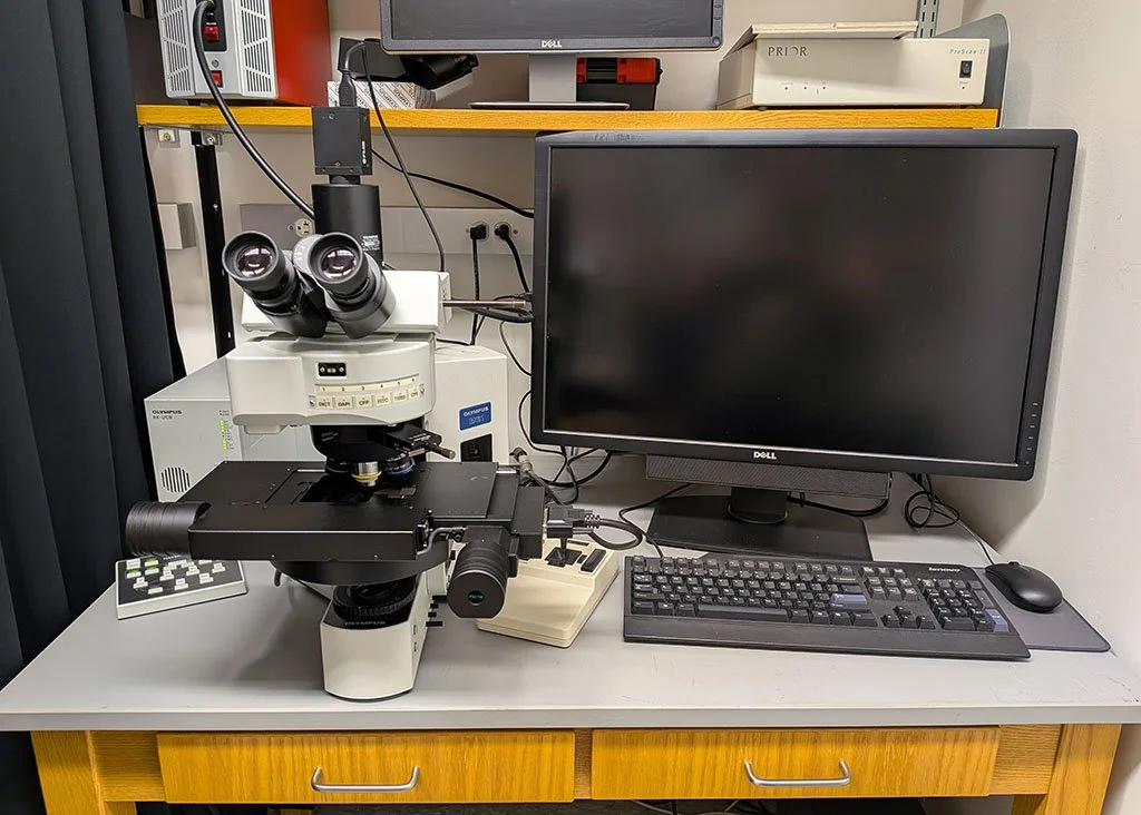
Olympus BX61WI
Microscope Type: Fully motorized upright fluorescence microscope for fixed tissue and slide-based imaging
Fluorescence Illumination: High-intensity Xenon arc lamp for broad-spectrum excitation across UV–visible wavelengths
Filter Sets: Interchangeable fluorescence filter cubes covering common fluorophores (DAPI, CFP, GFP, YFP, TRITC, Cy5)
Objectives: Wide range of high–numerical aperture plan achromat and plan apochromat objectives (4X, 5X, 10X, 20X, 40X, 63X (oil), 100X (oil)
Motorization:
Motorized stage for precise XY positioning, tiling, and multi-position imaging
Motorized filter turret and focus drive for automated acquisition workflows
Imaging Capabilities:
Multichannel fluorescence imaging
Z-stack acquisition for 3D reconstruction
Large-area image tiling and stitching
Camera System: High-sensitivity sCMOS camera for low-noise detection and rapid acquisition
Software Integration: Supports automated multidimensional imaging (XYZ, time, channels) and quantitative analysis workflows
Applications: Ideal for immunofluorescence imaging of tissue sections (including skin), fluorescent reporter analysis, histopathology, and high-resolution documentation of labeled specimens
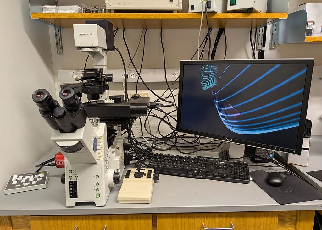
Olympus IX81
Microscope Type: Fully motorized inverted research microscope optimized for live-cell and in vitro imaging
Fluorescence Illumination: Metal halide fluorescence light source
Filter Sets: Interchangeable fluorescence filter cubes supporting common fluorophores (DAPI, CFP, FITC, RFP, TXRED, Cy5)
Objectives: Broad range of plan achromat and plan apochromat objectives (4x, 5x, 10x, 20X, 40X, 63x (oil)
Motorization:
Motorized XY stage for multi-position imaging, tiling, and automated scanning
Motorized Z-drive for precise focus control and z-stack acquisition
Motorized filter wheels/turrets for rapid channel switching
Imaging Capabilities:
Multichannel fluorescence imaging
Time-lapse imaging for dynamic cellular processes
Z-stacks for 3D reconstruction
Large-area image tiling and stitching
Camera System: High-sensitivity sCMOS camera for fast acquisition and low-light imaging
Environmental Compatibility: Fully compatible with stage-top incubation systems for temperature, CO₂, and humidity control
Software Integration: Supports multidimensional acquisition (XYZ, time, channels) and automated workflows
Applications: Ideal for live-cell imaging of keratinocytes, fibroblasts, and other skin-resident cells, as well as in vitro assays, migration studies, and fluorescence-based functional imaging