Bioimaging Core
-
Our core facility provides versatile stereo and macro zoom microscopy platforms designed for rapid, high-quality imaging of specimens at mesoscopic scales, bridging the gap between whole-organ visualization and cellular-level analysis. The Olympus SZ61TR stereomicroscope offers reliable brightfield imaging with excellent depth perception, making it ideal for routine dissection, sample preparation, and documentation of intact tissues, embryos, and small organisms. Its ergonomic design and straightforward operation enable efficient workflows for both novice and experienced users.
For more advanced applications, the Olympus MVX10 macro zoom microscope extends these capabilities by incorporating high-sensitivity fluorescence imaging and a CMOS camera for image and video aquisition. With a wide zoom range and superior optical performance, the MVX10 enables seamless transitions from low-magnification overview imaging to higher-resolution interrogation of specific regions of interest. This system is particularly well-suited for imaging fluorescent reporters in whole mounts, large tissue sections, and cleared samples, while preserving spatial context.
-
Our core facility offers a robust upright imaging platform centered on the Olympus BX51, configured for high-quality brightfield, phase contrast, and differential interference contrast (DIC) microscopy. This system provides flexible, high-contrast visualization of a wide range of biological specimens, making it an essential tool for routine histological analysis and live cell imaging. Equipped with a high-resolution color CMOS camera, the BX51 supports high resolution image acquisition and documentation of experimental observations.
For investigators within the Skin Biology and Diseases Resource-Based Center, this platform is particularly valuable for examining standard histological preparations such as H&E-stained sections, as well as assessing epidermal architecture, dermal organization, and adnexal structures. Phase contrast imaging facilitates the study of cultured keratinocytes, fibroblasts, and other skin-resident cell types without the need for labeling, while DIC provides enhanced contrast for visualizing fine structural details in both live and fixed samples.
-
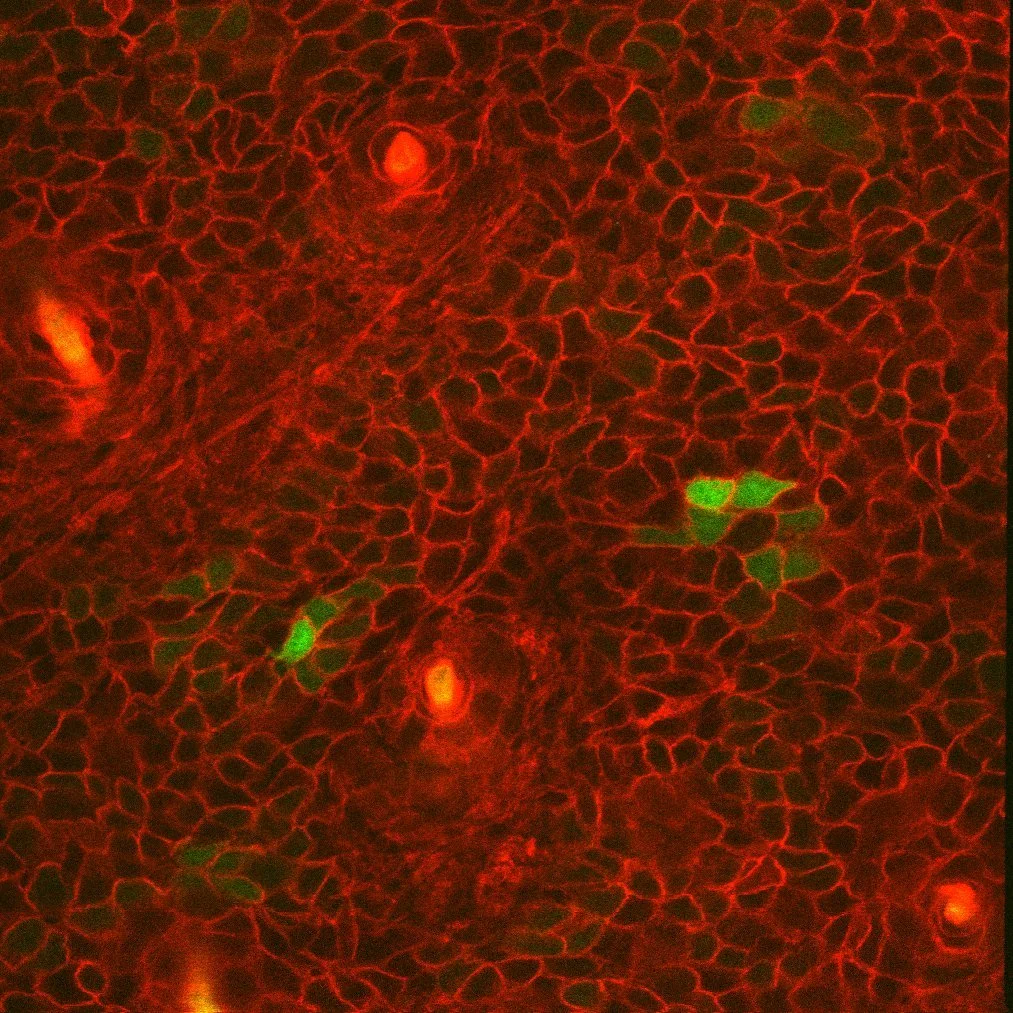
Our core facility provides flexible widefield fluorescence imaging through two fully motorized research platforms: the Olympus BX61 and the Olympus IX81. Together, these systems support a broad spectrum of applications ranging from fixed tissue imaging to live-cell dynamics, accommodating diverse experimental needs across multiple biological scales.
The upright BX61 is equipped with a high-intensity Xenon arc lamp and a comprehensive filter set spanning commonly used fluorophores, enabling sensitive detection across a wide spectral range. This configuration is particularly well-suited for imaging tissue sections, including immunofluorescence labeling of skin biopsies, where preservation of spatial architecture is critical. The inverted IX81 platform is optimized for brightfield and fluorescence live-cell imaging and in vitro assays, providing consistent illumination and minimal phototoxicity.
Both systems feature precision motorized stages that enable automated image tiling and stitching, facilitating large-area imaging of specimens such as full-thickness skin sections or cultured cell monolayers. High-sensitivity sCMOS cameras support rapid image acquisition, multidimensional imaging (including z-stacks and time-lapse), and high-quality video recording.
-
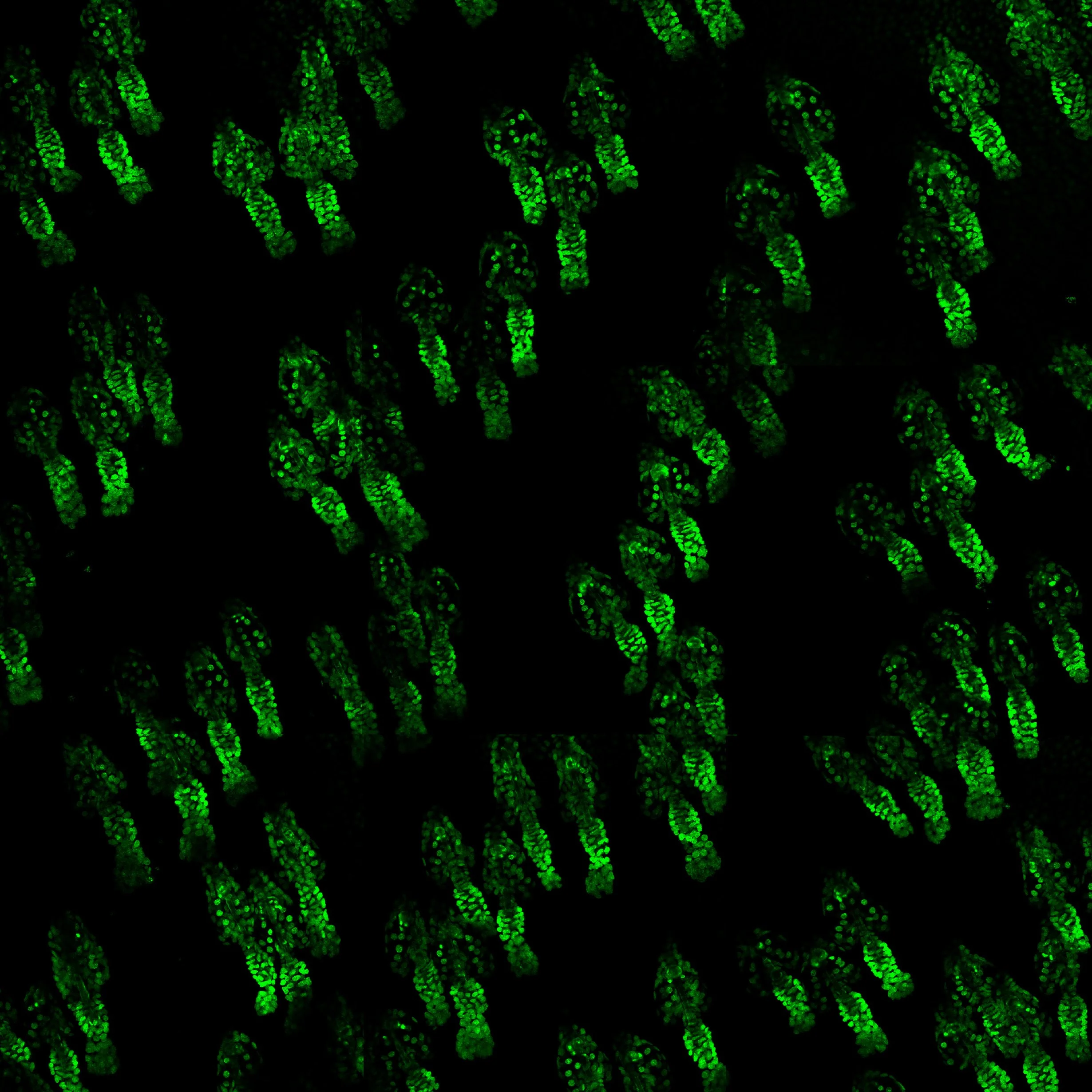
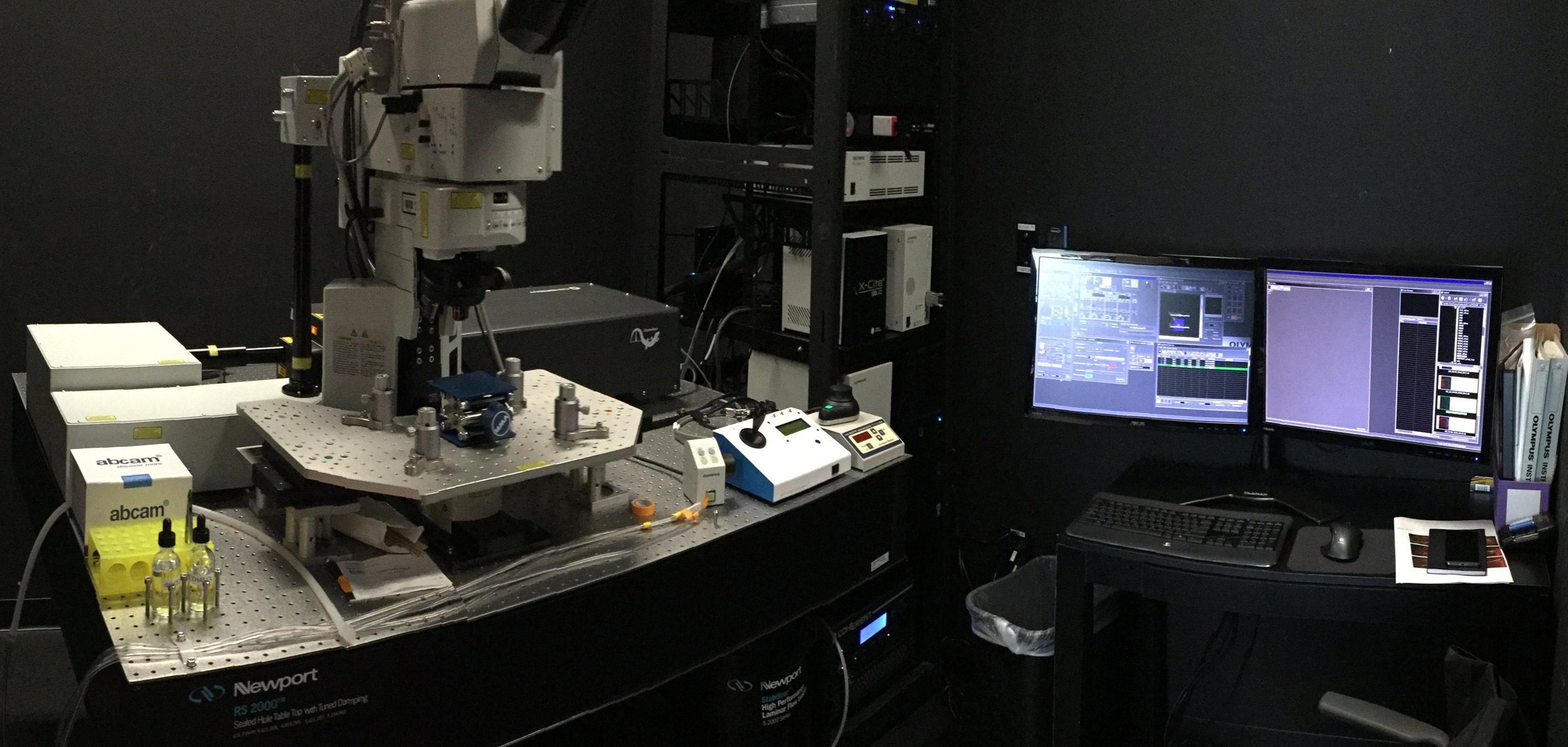
Our core facility provides state-of-the-art multiphoton imaging capabilities through two advanced platforms: the Olympus FV1200MPE and the next-generation Evident FV5000MPE. These systems enable deep tissue imaging with subcellular resolution, minimal phototoxicity, and intrinsic optical sectioning, making them ideally suited for live imaging of intact biological systems. These platforms are particularly powerful for applications in skin and ocular biology, where non-invasive intravital imaging allows longitudinal tracking of cells and structures in their native microenvironment. They support second harmonic generation (SHG) imaging of collagen, visualization of vascular and immune dynamics, and functional imaging approaches such as calcium signaling.
The FV1200MPE is equipped with a Coherent Chameleon Vision II laser, providing tunable excitation across a broad near-infrared spectrum for efficient multiphoton excitation of a wide range of fluorophores. Complementing this, the FV5000MPE integrates the cutting-edge Coherent Discovery NX dual-laser system, enabling simultaneous or sequential excitation at two independent wavelengths. This configuration significantly expands multiplexing capabilities and enhances imaging depth and signal quality, particularly in highly scattering tissues.
-
Our core facility provides flexible environmental control solutions to support high-quality live cell imaging across multiple microscopy platforms, including the Olympus IX81, Olympus MVX10, and our multiphoton systems. These setups enable stable, physiologically relevant conditions for both short- and long-term imaging experiments.
We offer the Bioptechs FCS3 environmental chamber, which features a heated, temperature-controlled stage and an integrated peristaltic pump for continuous perfusion of culture media. This system is particularly well-suited for experiments requiring rapid media exchange, pharmacological perturbations, or precise control of extracellular conditions during imaging.
In addition, the ibidi Stage Top Incubator provides comprehensive environmental regulation, including CO₂ control via a gas mixer, temperature stability, and humidity control. This platform supports extended time-lapse imaging of sensitive cell types by maintaining optimal culture conditions over prolonged periods.
Together, these systems enable dynamic imaging of cellular behaviors such as migration, proliferation, and signaling in real time. Their compatibility with multiple imaging modalities ensures seamless integration into diverse experimental workflows, from standard real-time live cell imaging to advanced multiphoton applications.
-
Our core facility offers advanced capabilities for whole-animal intravital imaging, enabling direct visualization of cellular and tissue dynamics within living organisms over time. These approaches are primarily supported by our multiphoton platforms, including the Olympus FV1200MPE and FV5000MPE, which provide deep tissue penetration, reduced phototoxicity, and high-resolution optical sectioning in intact specimens.
We have developed specialized workflows and instrumentation to support stable, reproducible imaging in live mice, including anesthesia delivery systems (isoflurane-based), temperature-controlled stages, and custom stabilization platforms for skin, ocular surface, and other accessible tissues. These configurations allow longitudinal imaging of the same region over hours to months, facilitating studies of stem cell behavior, tissue regeneration, immune responses, and disease progression in their native physiological context.
Particularly for skin and ocular surface biology, intravital imaging enables real-time observation of epithelial dynamics, neuronal activity, and vascular interactions with minimal perturbation. The systems also support functional imaging modalities, including calcium dynamics and label-free approaches such as second harmonic generation (SHG) for collagen visualization.
This unique capability positions the core facility at the forefront of in vivo imaging, empowering investigators to connect molecular and cellular processes with organism-level physiology in health and disease.
More info…
Core Director
Technical Director

Pantelis Rompolas PhD
Sixia Huang PhD